28Sep
CT, X-Ray or Ultrasound? A Simple Guide to Choosing the Right Scan
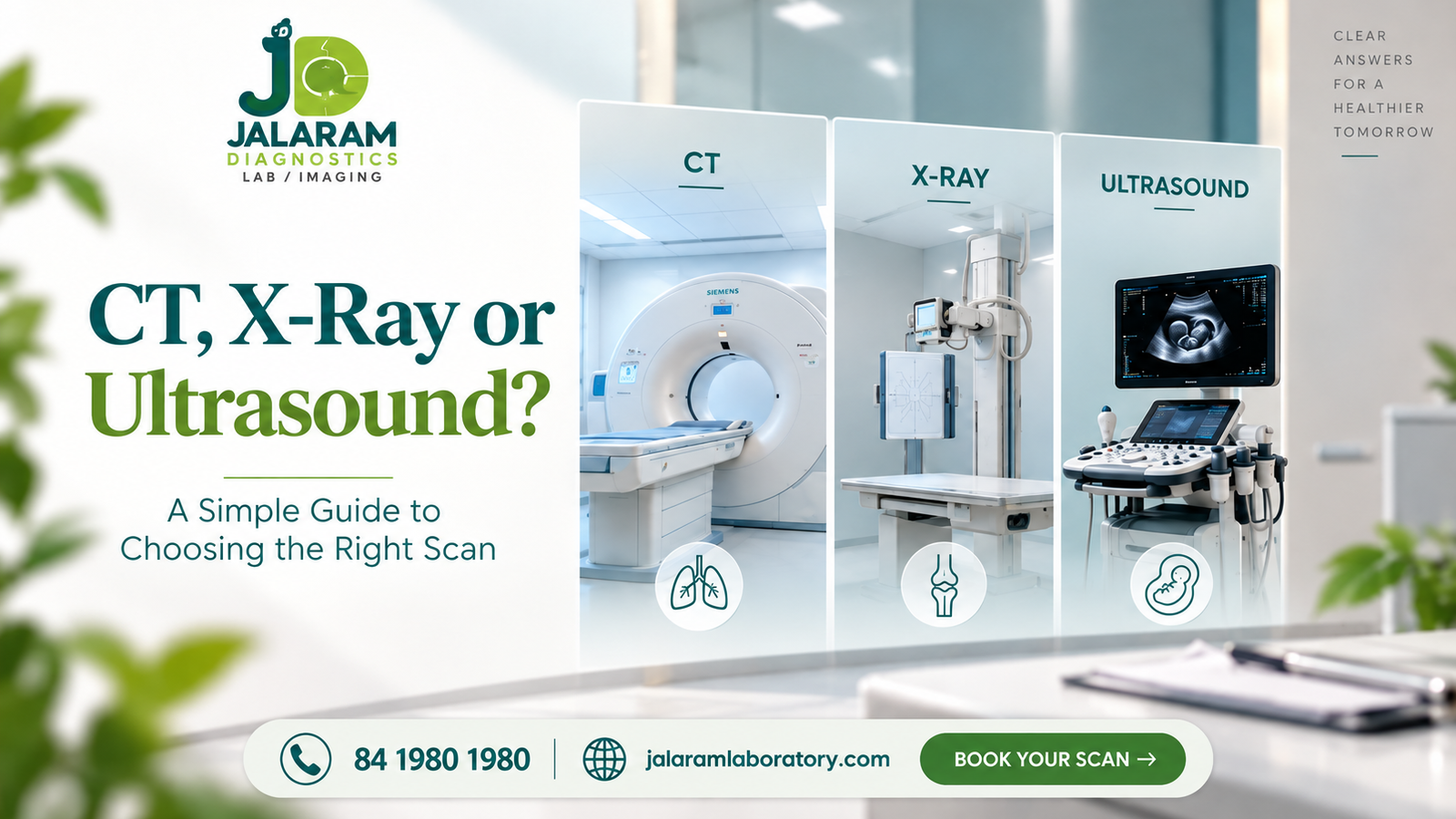
CT, X-Ray or Ultrasound? A Simple Guide to Choosing the Right Scan
When a doctor recommends an imaging test, it is natural to wonder why a particular scan has been advised and what it can show. CT scans, X-rays and ultrasounds are all commonly used to create images of structures inside the body, but each uses a different technology and serves different diagnostic purposes.
Understanding the basic differences between these imaging tests can help you feel more prepared and know what to expect when your healthcare professional recommends an investigation.
What Is Medical Imaging?
Medical imaging refers to diagnostic techniques that create images of structures inside the body. These images can help healthcare professionals examine areas such as bones, organs, soft tissues and blood vessels.
There is no single imaging test that is suitable for every situation. The choice depends on several factors, including the body part being examined, the suspected condition, urgency, age and medical history.
For this reason, the appropriate imaging investigation should always be determined by a qualified healthcare professional based on the individual clinical requirement.
CT Scan vs X-Ray vs Ultrasound
Although these investigations are all used for medical imaging, they work differently and provide different types of information.
X-Ray
An X-ray is one of the most commonly used imaging investigations. It uses a small amount of ionising radiation to produce images, particularly of bones and certain areas of the chest.
Doctors may recommend an X-ray when evaluating:
- Bone injuries and fractures
- Certain joint problems
- Chest-related concerns
- Other conditions where X-ray imaging is appropriate
One of the advantages of an X-ray is that the examination is generally quick and widely available, making it a commonly used first-line imaging investigation in appropriate situations.
CT Scan
A CT, or Computed Tomography, scan uses X-rays along with computer processing to create detailed cross-sectional images of the body.
Compared with a standard X-ray, a CT scan can provide more detailed information about internal structures. Depending on the clinical requirement, it may be used to examine areas such as the head, chest, abdomen and bones.
Some CT examinations may require contrast material to provide additional information in the images. Whether contrast is needed depends on the examination and will be determined by the healthcare team. Patients should follow the preparation instructions provided before the scan.
Ultrasound
Ultrasound uses high-frequency sound waves to produce real-time images of structures inside the body. Unlike X-rays and CT scans, ultrasound does not use ionising radiation.
It is commonly used to examine areas such as the abdomen, pelvis, thyroid and soft tissues. Ultrasound is also widely used during pregnancy.
Another application is Doppler imaging, which can be used to assess blood flow when clinically appropriate.
Which Scan Is Right for You?
There is no universally “best” imaging test. Each investigation has its own strengths, and the appropriate choice depends on what your doctor needs to evaluate.
| Imaging Test | Common Strength |
|---|---|
| X-Ray | Bones and chest imaging |
| CT Scan | Detailed cross-sectional imaging |
| Ultrasound | Soft tissues, organs and real-time imaging |
In some situations, one investigation may provide the information required. In others, additional imaging may be recommended to evaluate a particular concern more closely.
The important point is to follow the imaging recommendation provided by your healthcare professional rather than choosing a scan based only on which technology appears more advanced.
Why Professional Imaging Matters
The usefulness of an imaging investigation depends on more than simply having a scan performed. Appropriate technique, suitable equipment, trained professionals and proper interpretation all contribute to the quality of the diagnostic process.
If you have been advised to undergo a CT scan, X-ray or ultrasound, make sure you understand the preparation instructions provided by your healthcare team. Depending on the specific examination, you may be given instructions related to fasting, preparation or other precautions.
Following these instructions can help ensure that the examination is performed appropriately.
Frequently Asked Questions
Is an ultrasound better than an X-ray?
Neither is universally better. Ultrasound and X-ray use different technologies and are suitable for different diagnostic questions. The appropriate investigation depends on the area being examined and what your healthcare professional needs to evaluate.
Is a CT scan the same as an X-ray?
No. Both use X-rays, but a CT scan combines X-ray imaging with computer processing to create detailed cross-sectional images. A standard X-ray generally provides a simpler image and is commonly used for areas such as bones and the chest.
Do all imaging tests require preparation?
No. Preparation depends on the specific examination. Some ultrasound or CT studies may require particular instructions, while others may not. Always follow the instructions provided by your healthcare team.
How do I know which scan I need?
The appropriate scan depends on your symptoms, medical history, the body part being examined and what your doctor needs to evaluate. Your healthcare professional will recommend the investigation that best suits the clinical requirement.
Imaging Services at Jalaram Diagnostics
Jalaram Diagnostics provides a range of diagnostic and imaging services to support different healthcare needs. Depending on your doctor’s recommendation, investigations such as CT, X-ray and ultrasound can be used to evaluate different parts of the body.
The diagnostic services are supported by appropriate technology and professional teams, with a focus on organised patient care and reliable reporting.
If you have been advised to undergo an imaging investigation, understanding the basic purpose of the recommended scan can help you feel more prepared. Rather than choosing an imaging test on your own, discuss your requirements with your healthcare professional and follow the recommended investigation and preparation instructions.
Jalaram Diagnostics can support you with the required imaging investigation and help make the diagnostic process convenient and organised.